
Cell Culture
Optimised solutions for research
From experts for experts
Life sciences have a rule: Cell biology is not everything, but without cell biology, everything is nothing. As almost every scientific hypothesis, no matter how good, must firstly be proven to be correct in the smallest unit of life which is the cell. Cell culture is therefore indispensable in basic and drug research. Elucidating complex signal pathways, drug discovery and development, efficacy and toxicity studies, or reducing animal testing according to the 3R rules – It is almost impossible to do these without cell models. The number of cell lines and culture systems available at your disposal are enormous. Are you working with primary and secondary cells or cell lines, in suspension or adherent, 2D or 3D cultivation? The right choice is crucial for the success of your scientific work. What should you consider?
With SARSTEDT, you have a reliable partner at your side to answer all your questions. Our experienced experts will work with you to identify your specific requirements and put together the optimum solution from our extensive portfolio. With our high-quality culture vessels, bioreactors, storage & filtration systems, we have the right tools for all your tasks. Discover our innovative solutions and benefit from our expertise in cell biology.

Cells and cell lines: The spoiled for choice
The first and most important question is often: Which cell line is suitable for finding answers to your research question? Both primary cells and secondary cell lines can be valuable tools.
Primary cells are isolated directly from human or animal tissues, or body fluids. They provide more meaningful results than secondary cell lines, as they accurately reflect the morphological and physiological properties of the tissue from which they were taken. However, lifespan and proliferation of these cells is finite and slow, and cultivation is often more time-consuming and complex.
Secondary cell lines such as HeLa, HEK-293, or CHO cells are, by contrast, capable of division and survival (immortalised). They can be easily cultivated using established protocols, divide faster, and are commercially available for a relatively low price. However, they do not accurately reflect the physiological properties of the original tissue as precisely as primary cells.
Once you have selected the appropriate cells or cell lines for your research question, it is worth checking a few points:
Authenticity: Are you actually working with the correct cell line or cell type?
Contamination: Is the culture free of mycoplasmas, viruses, yeasts or cross-contamination from other cells?
Low passages: Immortalised cell lines, e.g. tumour cells, often exhibit high genetic instability and can degenerate after frequent passaging - is the number of passages therefore low?
Culture conditions: Everything needs to be correct!
Regardless of whether you are working with primary cells or secondary cell lines, specific culture conditions play a crucial role. This starts with choosing the optimal growth surface in your culture vessel .
Cells that adhere to surfaces have different needs than cells cultivated in suspension.
Hydrophilic culture vessels provide optimal culture substrate for adherent cells. They facilitate the initial adsorption and subsequent adhesion of cells.
Hydrophilic culture vessels with additional polar groups facilitate the adsorption and adhesion of primary cells or sensitive cell lines.
Hydrophobic culture vessels, on the other hand, reduce the adhesionof suspension cells and keep them in the solution.
Especially with adherent cells, the surface of the culture vessel must be completely flat so that your cells can form a uniform monolayer and do not accumulate at the edges of the vessel.
Depending on the application, different culture vessels are available for this purpose:
Flasks: For high cell counts, high protection against contamination by screw cap
Dishes: For moderate cell counts
Plates: For low cell counts, e.g. for running experiments in parallel
In addition to the appropriate culture vessel, culture media, environmental factors and the human factor play a crucial role.

3D cell culture: It's going round!
Classic 2D cell cultures are suitable for many basic research questions. However, results from a monolayer of identical cells can only be applied to tissues to a very limited extent, let alone an organ or even an entire organism.
To get as close as possible to the organ complexity, spheroid cultures play an increasingly important role, because:
They form cell-cell- and cell-matrix contacts.
They can be composed of different cell types.
Their 3D structure creates nutrient, oxygen, or active substance gradients.
They close the gap between the in vitro and in vivo situation.
They can help reduce animal testing.

BIOFLOATTM: Fast and reproducible spheroid culture
Ideally, uniform spheroids can be generated in a quick and reproducible manner - even when dealing with challenging cell lines. The appropriate cell culture plate can make your work significantly easier and lead to measurably better results.
The robust and highly anti-adhesive BIOFLOATTM surface ensures that adherent cells preferentially form cell-cell contacts. Compared to other anti-adhesive surfaces this means the following, for your 3D culture:
Faster spheroid formation after 2 to 24 hours depending on the cell line
Higher reproducibility due to highly uniform and round spheroids
Reliable spheroid formation, even with challenging cell lines
As a 96 well cell culture plate in ANSI/SLAS standard dimensions , BIOFLOATTM plates are suitable for automated high-throughput procedures, such as in the preclinical phase of drug research, in toxicological studies, and in cancer research .

Product Highlights
TC inserts for complex experiments in cell and tissue culture
In addition to spheroid culture, in vivo conditions can also be simulated by using cell culture plates and corresponding TC inserts :
The 2-compartment system provides your cells with an environment similar to an in vivo situation.
The inserts are equipped with an ultra-thin, microporous membrane that allows for optimal cell adhesion.
The 2-compartment system allows you to conduct complex experiments, such as transport, secretion and diffusion studies, co-cultivations, 3D cell cultures like organotypic skin models and more.
Cell spotlight: Products for cell microscopy
A milky film at the bottom of the culture vessel and a cloudy culture medium are all that can be seen with the naked eye. Mammalian cells measure between 1 µm and 30 µm, with human cells averaging around 25 µm. The condition or growth of your cell culture can therefore only be assessed by light microscopy. Further details, e.g. specific organelles, surface molecules or the expression of certain gene products, can be revealed only after appropriate staining or labelling, for example by using a fluorescence microscope.

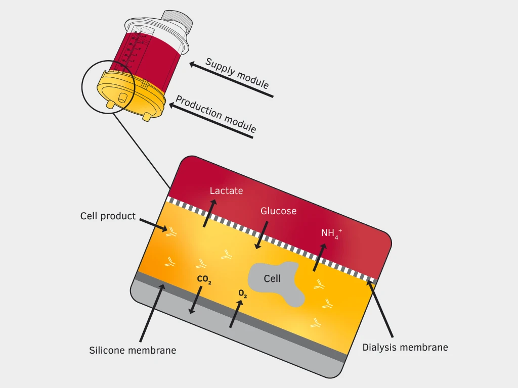
The cell as a production plant: miniPERM® bioreactor
Producing biomass or cell products such as proteins effectively and in larger quantities is difficult in conventional culture flasks because in addition to a lot of reactions, it also requires the necessary space in the incubator.
Instead, the use of a bioreactor, which constantly supplies your cells with medium, removes metabolism end products and ensures the highest possible yield, is recommended.
On a laboratory scale, we do not just want a high yield, but also easy handling. For these purposes, production in the miniPERM® bioreactor is ideal.
Optimum design: The 400 ml nutrient module is separated from the 35 ml production module by a dialysis membrane for effective nutrient supply to your cells. Meanwhile, your product (< 12.5 kD) remains in the production module.
High cell density: Up to 1x10⁷ cells/ml density is possible, which is equivalent to approximately 20 large culture flasks or three 1litre roller bottles.
Optimal gas exchange: The outside of the production module is made of an O2- and CO2-permeable silicone membrane.
Versatile: A wide variety of mammalian, plant and insect cells can be cultivated.
Compact and simple: Using a universal turning device, up to four bioreactors can be compactly accommodated in the incubator.
Product Highlights
Troubleshooting: You can avoid these stumbling blocks
Despite all care and experience, sometimes problems can arise. Hereare the typical problems and how to avoid them:
Pipetting: Pipetting quickly puts stress on your cells and, in the worst case, they can be destroyed by shear forces. When changing media, the pipette tip should not touch the bottom of the culture vessel to avoid damaging the cell layer.
Centrifuging: Centrifuge your cells only as long and as fast as necessary to avoid unnecessary stressing of the cells.
Circular cell growth: Possible causes for this can be vibrations in the environment. Is your incubator next to an old fridge? Do your colleagues regularly slam the doors? Is a neighbouring laboratory currently undergoing renovation?
No growth in the middle of the well: If the volume of liquid in the well is too low, the meniscus can push the cells to the edge of the well. If your cells get collected at the edge in high density, it can lead to contact inhibition and reduced or no further growth. Using sufficient quantities of culture medium will prevent this.
Mycoplasmas: These bacteria, invisible under light microscope, can significantly affect or alter the growth and metabolism of your cells. Due to their lack of a cell wall, they are resistant to many antibiotics commonly used in the laboratory, such as penicillin, streptomycin or gentamicin. They also pass through sterile filters with a pore size of up to 0.2 µm without any issue. Regular checks, e.g. with PCR-based tests, provide certainty and sterile work routine reduces the contamination risk.
Cross-contamination and authenticity: Is your cell line the one you think it is? This is definitely a question worth asking, as cross-contamination occurs in laboratories where a variety of cell lines are used. Before and after a project, it is therefore worth taking a closer look at your own cell culture.
Cryopreservation: Store cell materials correctly.
Where do you usually get your primary cells or cell lines from? Presumably from the freezer or a nitrogen tank. Cryopreservation is essential in cell biology, as many cell lines are project-specific and therefore only needed temporarily.
Continuous cultivation is not feasible for a number of reasons. Time, material and the space in the incubator can be better used elsewhere. In addition, with each passage, the risk of your cells entering senescence or developing genetic or morphological changes increases. A carefully maintained and stored cell bank also protects you from losing entire cell lines due to contamination, device failures or handling errors.
Cell storage: There is (almost) no such thing as too cold
In many laboratories, cells are stored, at least temporarily, in an ultra-low temperature refrigerator at around -80 °C. This is unsuitable for the permanent storage of your cells as intracellular ice formation and recrystallization can lead to rapid viability loss.
With storage at less than < -130 °C in the gas phase of your nitrogen tank, you are on the safe side, but you need suitable consumables that can withstand these temperatures. This is because beyond -80 °C, conventional polypropylene or polycarbonate containers are prone to material failure.
The CryoPure freezing system offers suitable materials, with its tubes, racks and boxes specially designed for these temperatures.
Cold resistant down to < -196 °C: CryoPure tubes, racks and boxes are optimised for extremely cold temperatures.
Ergonomic: Easy opening of the screw cap with just a single turn, easy handling, thanks to the special base design, colour coding with coloured caps and colour-coded inserts.

Product Highlights
Filtration in cell culture: Is everything sterile?
The autoclave and safety cabinet ensure sterile materials and working conditions for your cell culture work. However, the source of life for your cells should also be as sterile as possible, which is the culture medium. Sterilisation in an autoclave is not possible for heat-sensitive components. However, sterile filtration gives you the option to safely remove fungi, mycoplasma and other bacteria.
Suspended matter and larger particles can usually be removed with a 0.45 µm filter, but a 0.22 µm filter should be used for bacteria or fungi. If you want to be on the safe side regarding mycoplasmas, it is best to use a pore size of 0.1 µm. Due to their lack of cell wall, small pests are so malleable that they can easily pass through a 0.2 µm filter.

Product Highlights
Purity levels in cell culture: Keep it clean!
Time, money, energy, dedication and a lot of tolerance - those who work in science usually need all of these. Almost every researcher knows the feeling of identifying something wrong by taking a first look through the microscope. What are supposed to be adherent cells are floating dead in the medium or something is growing that shouldn't be growing there.
Trial and error, successes and failures are part of scientific work just like a pipette in the hand or a microscope on the table. Unnecessary setbacks can be avoided by keeping an eye on the purity levels of your materials from the beginning of your cell culture work.
Various factors play a role here:
Sterility: Guarantees that no viable bacteria or fungi are introduced into your culture.
Non-pyrogenic/endotoxin-free: Pro-inflammatorysubstances can remain on the material even if no viable bacteria are present.
Non-cytotoxic: For vital cell growth, no cytotoxic substances may enter your culture.
DNA-free: Preserves the authenticity of your cell line, as under certain conditions foreign DNA can be taken up from the medium by the cells and integrated into the genome.
TC Tested: Products with this purity level do not just meet the above requirements, they are free from DNase and RNase as well.
Get an overview of our purity levels and SARSTEDT QUALITY .
Life is not always science.
But science is our Life.
At SARSTEDT, our work follows the principle: Impress with quality, retain with service. Reliability and experience, innovation and dynamism – we build on proven and reliable values, combined with active processes and partnership-based thinking and action.
